About Flourescence
It took many years - since the discovery by Osamu Shimomura of the first proteins aequorin and green fluorescent protein (GFP) in the bioluminescent jellyfish Aequorea Victoria around the sixties - to develop a specific fluorescence microscope for live-cell imaging which permits essential progress in medical and biological sciences. Today it has become an important tool in Life and Materials Sciences.
INTRODUCTION TO FLUORESCENCE MICROSCOPY
Fluorescence is the phenomenon of subsequent specific emission of photons by a fluorophore (chemical compound) after the absorption of specific excitation photons. Fluorophores are used as a dye for staining cells, tissues or materials that can be observed as markers under a fluorescence microscope.
DESIGN OF A FLUORESCENCE MICROSCOPE
The basic function of a fluorescence microscope is to bring specific "excitation" wavelengths to a stained specimen (with a specific fluorophore) and to catch the subsequent weak emission wavelengths in order to visualize the fluorescent structures on the retina of the eyes or on an imaging camera.


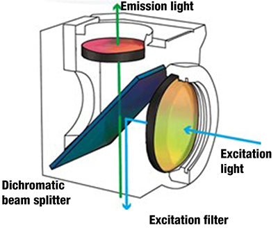
The excitation light of a microscope for fluorescence is provided with either a mercury vapor light source or a monochrome LED. The selection of the specific fluorophore excitation light is done by a so-called emission interference filter that is mounted in a cube together with a dichroic mirror (beam splitter) and the emission filter. The filtered excitation light is further reflected by the dichroic mirror through the objectives towards the sample.
The fluorophore(s) inside the specimen will absorb this excitation energy with some efficiency (quantum yield) and will start re-emitting the so-called emission energy, visible light with longer wavelengths than the absorbed light.
The emission light from the specimen that fluoresces will be collected by the objective, pass the dichroic mirror and is separated from excitation and other unwanted light wavelengths by the emission filter. The filtered emission light is further projected to the eyepieces or/and camera imaging sensor.

